Liquid chromatography tandem mass spectrometry
A chromatographic separation feeding a mass spectrometer, so a component can be weighed rather than only timed.
- Cited on certificates as
- LC-MS/MS
- Standard
- USP <736> Mass Spectrometry
- Reports
- Identification (MS) · Confirmation of identity
What LC-MS/MS is
Liquid chromatography tandem mass spectrometry puts two instruments in series. The first is the chromatography described on the HPLC page: it separates the mixture so components arrive at the second instrument one at a time rather than together.
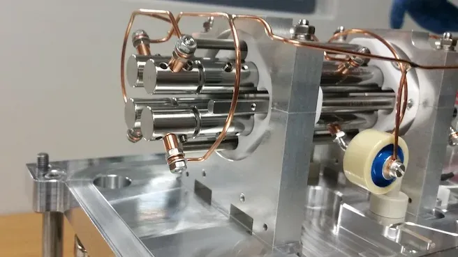
The second is a mass spectrometer, and “tandem” means it contains two mass analysers with a collision cell between them. The first analyser selects an ion by its mass-to-charge ratio, the collision cell breaks it apart, and the second analyser records the fragments. A compound is therefore characterised twice: by the mass of the whole and by the pattern it breaks into.
The measurement that matters is usually the first of those: the observed molecular weight against the value calculated from the declared structure — from a sequence for a peptide, from a formula for a small molecule.
As stated on a certificateChromatographic separation followed by tandem mass analysis, confirming the observed molecular weight against the theoretical value for the declared sequence.
How it works
Separation
The LC front end is the same technique described on the HPLC page. Its job here is to deliver one component at a time, because a mass spectrum of an unseparated mixture is a spectrum of everything at once.
Ionisation
Electrospray is the usual source. The eluent is sprayed through a needle held at high voltage, producing charged droplets that shrink until the analyte is left as a gas-phase ion. It is gentle enough to leave a large or fragile molecule intact — a peptide, a protein, anything that a harder source would take apart before it was weighed — which is why it is the one used here.
Precursor selection
The first analyser is set to pass one mass-to-charge ratio and reject everything else. What reaches the collision cell is therefore one species out of the whole eluent.
Fragmentation
The selected ion collides with an inert gas and breaks, and where it breaks is characteristic of the molecule. A peptide gives way at its amide bonds, so its fragments form a ladder whose spacings are residue masses; a small molecule loses identifiable groups instead. Either way the pattern, not just the mass, is the evidence.
Fragment analysis
The second analyser scans the fragments. The pattern is characteristic, which is what turns a mass into evidence about a structure rather than only a number.
Reading what it produces
- A large molecule in electrospray usually appears several times over, carrying different numbers of protons: a molecule of mass M shows up at (M + nH)/n for a series of n. Peptides and proteins do this readily; the spectrum looks busier than the sample is.
- Deconvolution folds that series back into one neutral mass. The number a certificate carries is that neutral mass, not a raw m/z.
- Agreement is judged against a tolerance, not exactly. Certificates issued from this method state the tolerance alongside the observed and theoretical values.
- A fragment spectrum reads as spacings rather than peaks: the gaps between adjacent fragments are residue masses, and it is the gaps that carry the sequence information.
What we report from it
Every one of these is a measurement made in this laboratory on the sample as received. The certificate names the method behind each figure, because the method is most of what the figure is worth.
- Identification (MS)
- The observed molecular weight against the theoretical value for the declared sequence, with the tolerance the method allows. A certificate states both figures rather than only the verdict, so a reader can see how close the agreement was.
- Confirmation of identity
- Where fragment ions are assigned, they support the identity beyond the intact mass alone. What a certificate can state depends on which transitions the method monitored, so it states them.
The instrument
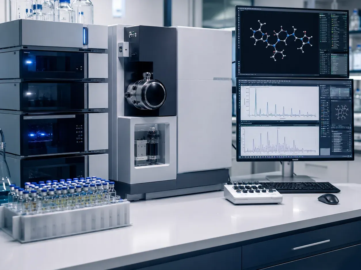
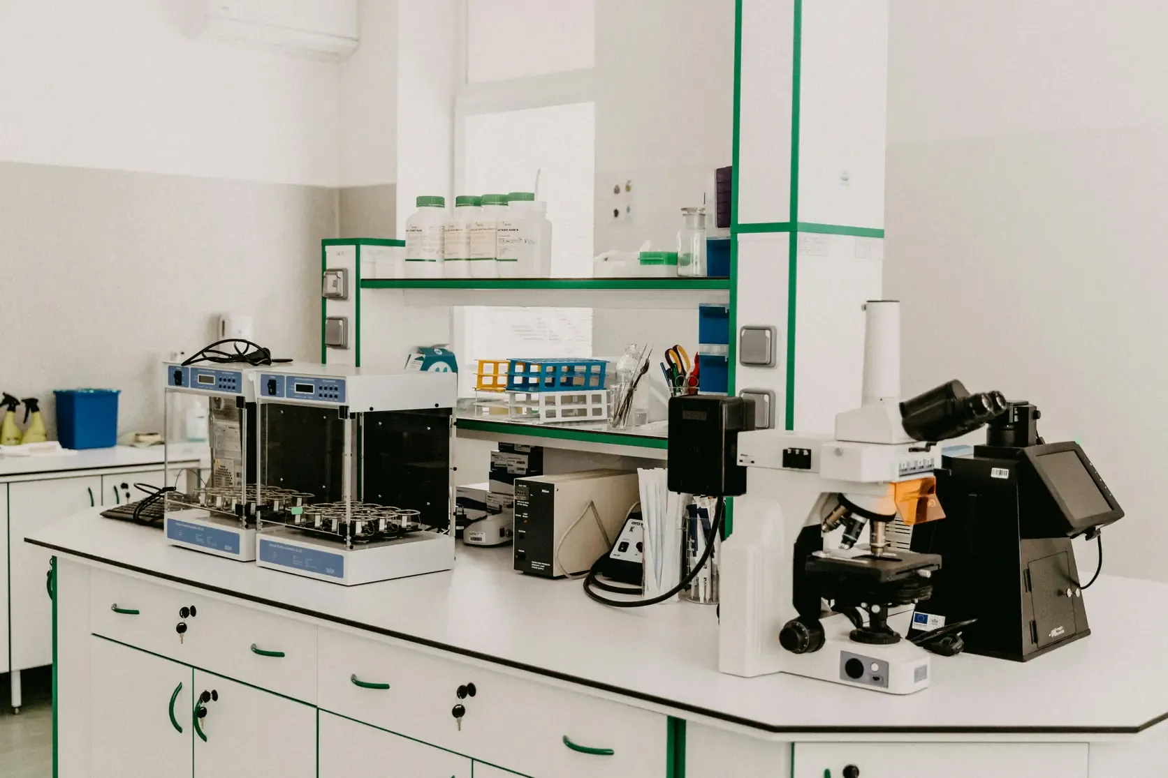
What a laboratory running this method has on the bench, and what each part decides about the result.
- LC front end
- Delivers components separately. Without it the spectrometer sees the mixture all at once.
- Electrospray source
- Turns dissolved molecules into gas-phase ions without breaking them.
- First mass analyser
- Selects one precursor mass-to-charge ratio out of everything arriving.
- Collision cell
- Fragments the selected ion against an inert gas at a controlled energy.
- Second mass analyser
- Records the fragments. A triple quadrupole scans a set of transitions; a time-of-flight analyser records the whole spectrum at higher resolution.
What it cannot establish
This list is the reason the rest of the page is worth reading. A figure, or an observation, means what its method can support and nothing past it.
- 01Mass is not structure. Two different molecules with the same formula have the same mass, and the intact mass alone cannot separate them. Distinguishing them needs the fragment pattern, and even then not always.
- 02A mass confirmation says nothing about how much is present. Ionisation efficiency depends on the compound and on what else is in the solution, so signal size is not proportional to quantity in the way a UV peak area is.
- 03Anything that does not ionise under the conditions used is not seen, whatever its concentration.
- 04It does not measure purity. A certificate’s purity figure comes from the chromatography, not from the mass spectrum.
- 05A result is only as good as the theoretical value it was compared against. The declared sequence has to be right for the comparison to mean anything.
Where it is used
- Confirming identity against a declared structure, sequence or formula
- Detecting deletion, insertion and oxidation variants by their mass difference
- Characterising an impurity a chromatogram has separated but not named
- Supporting an identity claim where retention time alone is not enough
Questions people arrive with
Asked about LC-MS/MS often enough to be worth answering here rather than by return of email.
Anything else, and the answer comes from a person rather than a page. Write to the laboratory.
Why does a certificate quote both an observed and a theoretical mass?
Because the verdict depends on the gap between them and on the tolerance allowed. Printing only “identity confirmed” asks the reader to take the judgement on trust; printing both figures lets them make it themselves.
Does a matching mass prove the compound is what it says?
It is strong evidence and not proof. Isomers share a mass, and so do some unrelated formulae. Fragment analysis narrows it further, and the certificate states which of the two was done.
Why is quantity not taken from the mass spectrum?
Response depends heavily on the compound and on what else is present in the solution — an effect called ion suppression. Chromatography with UV detection is more even-handed across components, so quantity is taken from there.
What is the difference between MS and MS/MS?
A single-stage instrument weighs what arrives. A tandem instrument weighs it, breaks it, and weighs the pieces — which is what lets a result say something about structure rather than only about mass.
Also cited on certificates
HPLC
The separation most purity figures begin as: a mixture pushed through a column, and a detector recording what comes off and when.
GC
The separation for things that will travel as a vapour — residual solvents above all, which is the line a purity figure by HPLC cannot fill in.
SFC
The separation that can tell two mirror images apart — and the only one here that can, because on every other column they are the same compound.
SEM
The method that produces a picture rather than a number — and the one whose result depends most on how the sample was chosen.
Holding a certificate that cites LC-MS/MS?
Check its number against our record. No account, and the same check is open to anyone holding the document.