Gas chromatography with flame ionisation detection
The separation for things that will travel as a vapour — residual solvents above all, which is the line a purity figure by HPLC cannot fill in.
- Cited on certificates as
- GC-FID
- Standard
- USP <467> Residual Solvents
- Reports
- Residual solvents · Volatile impurities · Assay of a volatile substance
What GC is
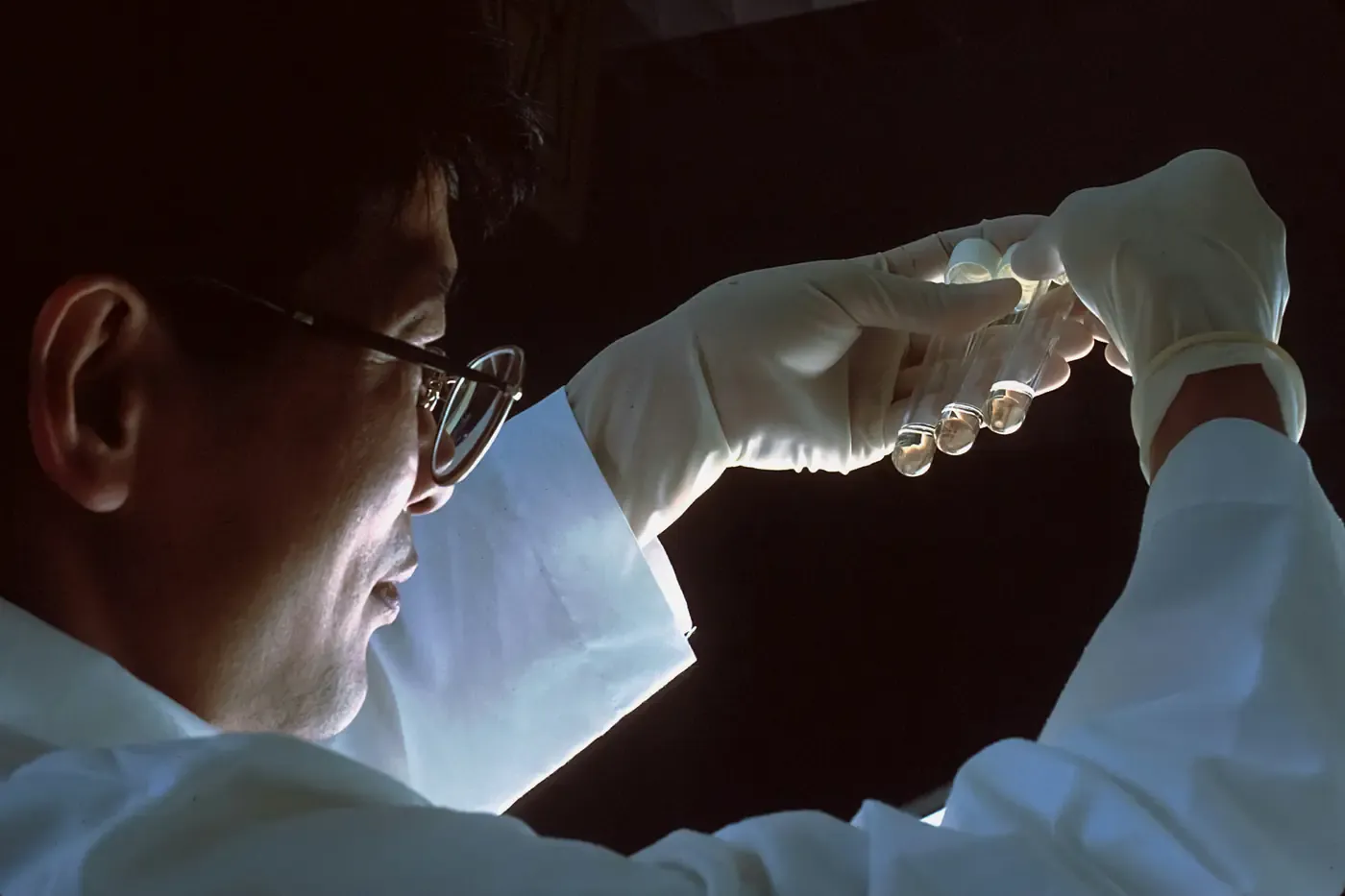
Gas chromatography separates a mixture in the vapour phase. The sample is vaporised at the head of a heated column, an inert carrier gas — helium, hydrogen or nitrogen — pushes it through, and each component is held back by the thin coating on the column wall for a different length of time. What leaves the far end is a series of separated compounds, each at its own retention time.
The condition is volatility. A compound has to survive being heated and has to travel as a gas, which is the whole difference from liquid chromatography: HPLC carries the sample dissolved, so it can analyse things that would decompose long before they boiled. Most actives are among them, which is why a purity figure normally comes from a liquid method and the residual solvent figure from this one.
“FID” is the detector. The eluent is burned in a hydrogen flame and the ions that combustion produces are collected as a current, which is very nearly proportional to the mass of carbon passing through. It responds to almost any organic compound, over about seven orders of magnitude, and it is destructive: whatever it measures is gone.
As stated on a certificateVolatile components are carried by an inert gas through a heated capillary column and separated by boiling point and by affinity for the column coating, then burned in a hydrogen flame whose ion current is proportional to the carbon passing through it.
How it works
Introduction
Either the sample itself is injected, or — for residual solvents — the sealed vial is warmed and the vapour above the sample is sampled instead. That second route is headspace injection, and it keeps a non-volatile matrix out of the column entirely rather than asking the column to cope with it.
The inlet
A heated inlet flashes the sample to vapour. Split injection sends a measured fraction onto the column and vents the rest, which prevents overload; splitless sends nearly all of it, which is what trace work needs. The split ratio is part of the method, because it decides what fraction of the sample the figure is about.
The oven programme
The column sits in an oven that is ramped rather than held. Starting cool separates the early, volatile peaks; heating drives the heavier ones off in reasonable time. The programme is part of the method in the way a gradient is for HPLC, and changing it moves every retention time on the chromatogram.
Separation
Components partition between the carrier gas and the stationary film coating the inside of the capillary. Volatility sets the broad order and the chemistry of the coating decides the rest — a polar column and a non-polar one will put the same two compounds in different orders.
Detection
The flame ionisation detector burns what arrives and measures the ion current. The area under each peak is proportional to the carbon that produced it, so quantitation is by area against a standard run under the same programme.
Reading what it produces
- The x-axis is time and the y-axis is detector response, exactly as in a liquid chromatogram. A retention time identifies a compound only against a standard run on the same column under the same oven programme; it is not a property of the compound alone.
- Peak area is proportional to carbon mass, not to moles and not to mass of compound. Two solvents present in equal amounts do not give equal peaks, so quantitation needs a response factor or a calibration for each one.
- A solvent peak from the injection itself sits early in almost every chromatogram. It is the sample diluent rather than a finding, and a chromatogram read without knowing which peak that is will over-report.
What we report from it
Every one of these is a measurement made in this laboratory on the sample as received. The certificate names the method behind each figure, because the method is most of what the figure is worth.
- Residual solvents
- Which solvents remain from synthesis or purification and how much of each, usually by headspace against the limits in USP <467>. This is the determination gas chromatography is most often on a certificate for.
- Volatile impurities
- Volatile organic compounds present alongside the declared substance, identified by retention time against standards and quantified by area.
- Assay of a volatile substance
- Where the substance itself is volatile, its content against a reference standard. It does not apply to an active that cannot be vaporised intact, which covers most of them.
The instrument
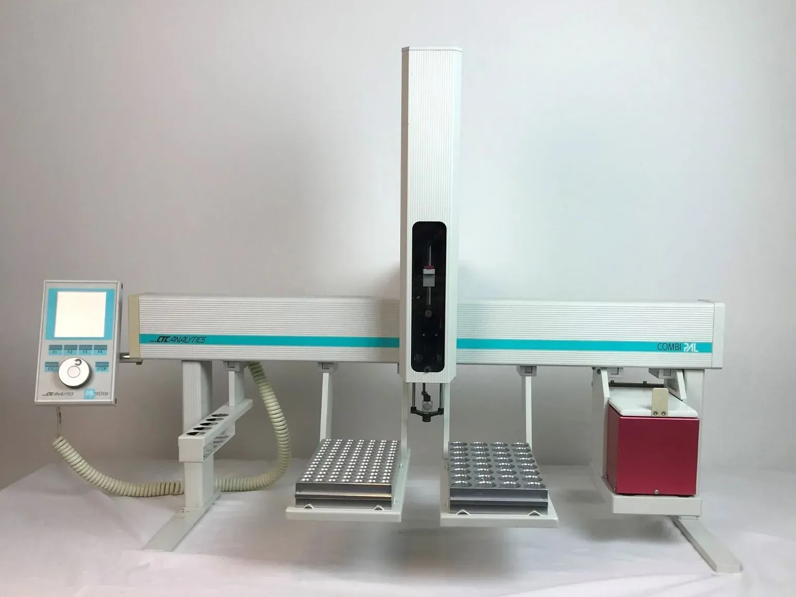
What a laboratory running this method has on the bench, and what each part decides about the result.
- Carrier gas supply
- Helium, hydrogen or nitrogen at a regulated flow. The gas is the mobile phase, and a drifting flow moves every retention time.
- Headspace sampler
- Warms the sealed vial and draws the vapour above the sample. It is what lets a non-volatile material be tested for volatile content without putting that material on the column.
- Heated inlet
- Flashes the injected volume to vapour, and splits it or does not. The split ratio decides what fraction of the sample reaches the column.
- Capillary column
- A long, narrow tube whose inner wall carries the stationary film. Length, bore and film chemistry set what can be resolved from what.
- Column oven
- Holds and ramps the temperature to a programme. Retention times are a function of that programme, so it is quoted with the method.
- Flame ionisation detector
- Burns the eluent in a hydrogen flame and measures the resulting ion current.
The detector decides what is visible
The column separates; the detector is what turns that separation into a number. It is also the half a method name mentions only in passing — the -UV in RP-HPLC-UV is not a suffix, it is the reason the purity figure is an area percentage at one wavelength. Every detector is blind to something, and what it cannot see does not appear on the chromatogram however much of it is there.
Flame ionisation
- Sees
- Almost any compound with carbon–hydrogen bonds, linearly over about seven orders of magnitude. It is the general-purpose detector and the one behind most residual solvent figures.
- Blind to
- Water and the permanent gases, and it responds poorly to heavily substituted carbons such as formic acid or carbon tetrachloride. It is also destructive: what it measures is burned, so nothing downstream can look at it again.
Thermal conductivity
- Sees
- Anything whose thermal conductivity differs from the carrier gas, which is everything — including the water and permanent gases that FID cannot see.
- Blind to
- Nothing in particular, and it pays for that with sensitivity: one to two orders of magnitude less than FID. It is chosen where universality matters more than trace detection.
Electron capture
- Sees
- Electronegative compounds, halogenated ones above all, far below the levels FID reaches. It is the detector behind organochlorine residue figures.
- Blind to
- Everything without an electron-capturing group, which is most organic chemistry. Its response is also strongly non-linear over a wide range, so a calibration has to bracket the expected level rather than span it.
Nitrogen–phosphorus
- Sees
- Nitrogen- and phosphorus-containing compounds selectively, which suppresses a hydrocarbon matrix that would otherwise bury them.
- Blind to
- Compounds containing neither element. The bead that gives it its selectivity is consumable and drifts as it ages, so it needs standardising more often than FID.
Flame photometric
- Sees
- Sulfur and phosphorus, by the light their combustion emits at element-specific wavelengths.
- Blind to
- Everything else. Its sulfur response is roughly quadratic with concentration rather than linear, which is a trap for anyone quantifying it as though it were not.
Mass spectrometry
- Sees
- Mass-to-charge ratio, and under electron ionisation a fragmentation pattern reproducible enough to match against a spectral library — so it can name a peak nothing else on this list can.
- Blind to
- Compounds that do not survive the inlet or the source. Response varies widely between compounds, so a tall peak beside a short one is not a statement about their relative amounts.
What it cannot establish
This list is the reason the rest of the page is worth reading. A figure, or an observation, means what its method can support and nothing past it.
- 01Anything not volatile is invisible, and most of what a certificate is about usually is not. Gas chromatography says nothing about the purity, identity or molecular weight of an active that will not travel as a gas; it is on a certificate for what the liquid methods cannot reach.
- 02A compound that decomposes at inlet or column temperature is reported as its decomposition products, or not at all. Thermally labile material has to be derivatised first, and a derivatisation that goes to ninety per cent completion yields a figure about the derivative rather than about the sample.
- 03FID area is proportional to carbon, not to substance. Without a response factor for each analyte, an area percentage across a chromatogram is not a composition.
- 04Water is not detected by FID at all, so a gas chromatogram carries no information about water content. That determination is Karl Fischer titration, and a certificate that needs it needs that analysis run.
- 05A headspace result describes what was in the vapour above the sample under the conditions used. Equilibration temperature, time and the matrix itself all change how much of a solvent partitions into that vapour, so the figure is comparable only against a calibration made the same way.
Where it is used
- Residual solvents from synthesis and purification, against the limits in USP <467>
- Volatile impurity profiles alongside a purity figure produced by a liquid method
- Solvent identity confirmation, where a process is expected to leave one and not another
- Volatile components of botanical extracts and essential oils
- Assay of substances that are themselves volatile
Questions people arrive with
Asked about GC often enough to be worth answering here rather than by return of email.
Anything else, and the answer comes from a person rather than a page. Write to the laboratory.
What does GC stand for in testing?
Gas chromatography: a separation in which the sample travels as a vapour, carried through a heated column by an inert gas and measured by a detector at the outlet. On an analytical certificate it is usually the method behind a residual solvent figure. Worth knowing that the same two letters mean something unrelated in clinical microbiology, where GC is the gonococcus and “GC/CT testing” screens for two infections. Nothing on this page concerns that.
Why is purity measured by HPLC and residual solvents by GC?
Because the two techniques reach different things. Most actives cannot be vaporised without being destroyed — peptides and proteins certainly, and any thermally labile molecule — so gas chromatography cannot measure their purity. A residual solvent is volatile and present in traces, which is precisely what a headspace GC method is built for. A certificate carrying both is not repeating itself; it is reporting two determinations that neither method could have made alone.
How does GC-MS quantify something, and what is a detection limit?
The chromatograph separates, the mass spectrometer measures mass-to-charge, and the amount is read from the area of a chosen ion against a calibration built from standards of known concentration. The limit of detection is the smallest amount distinguishable from noise; the limit of quantitation is the smallest that can be measured to a stated precision, and it is higher. A result reported below the limit of quantitation is evidence that something is present, not a measurement of how much.
Can gas chromatography be used on biological samples?
The technique can, where the analytes are volatile or can be derivatised to be. This laboratory does not: we issue certificates about materials rather than about people, and we ask that no clinical or personal health information be sent to us. A certificate here concerns one batch of one substance.
Is this method run at ZEIKKA?
Yes. Headspace gas chromatography with flame ionisation detection is run here, most often for residual solvent against the limits in USP <467>. The certificate names the method and the date, and reports which solvents were found and how much of each.
Also cited on certificates
HPLC
The separation most purity figures begin as: a mixture pushed through a column, and a detector recording what comes off and when.
LC-MS/MS
A chromatographic separation feeding a mass spectrometer, so a component can be weighed rather than only timed.
SFC
The separation that can tell two mirror images apart — and the only one here that can, because on every other column they are the same compound.
SEM
The method that produces a picture rather than a number — and the one whose result depends most on how the sample was chosen.
Holding a certificate that cites GC-FID?
Check its number against our record. No account, and the same check is open to anyone holding the document.